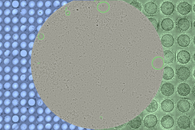
By combining cryo-electron microscopy (cryo-EM) samples are automatically imaged and by artificial intelligence (supervised machine learning) particles are automatically detected. The approach offers a valuable additon to currently existing analysis techniques that specifically impact purity assesment and characterization.
Extracellular Vesicles (EVs) are lipid membrane delimited particles released from all living cells. They contain proteins and genetic materials and play important roles in intercellular communication, making tem of broad interest for diagnostics and therapeutics.
The content, size and characteristics of EVs vary enourmously and not two EV particles are identical. Additionally, because EVs share overlapping physcial properties with other particles such as proteins, liprproteins and viruses it is difficult to obtain samples that are both highly concentrated and highly pure. While cryo-EM can directly visualize the lipid bilayer, a key characteristic for EVs, the aquisition and detection of EV particles is often performed manually, making the process slow and subjective.
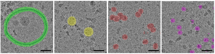
By combining automated cryo-EM image acquisition and supervised machine learning, and using models for the lipid bilayer to detect EVs, as well as several lipoprotein particles (LDL, VLDL, HDL) we were able to automaticlly detect hundreds of particles in thousands of images and determine their size distributions.
This approach enables fast and objectve detection of EVs and co-isolating particles, making it possible to assess the purity of samples and the size ditibution of partciles. These measurements might be omportant for diagnostic and therapeutic applications.
The results are published in Journal of Extracelular Vesicles: 10.1002/jev2.70273