Sex Chromosome Biology
Many species use sex chromosomes to determine sex and sex-specific gene expression. In mammals, females carry two X chromosomes while males carry an X and Y; since the Y carries under 70 genes and the X more than 1000, female X-linked expression would double male levels. Mammals equalize expression of X-linked genes by inactivating one X per female cell. XCI is controlled by the ~800kb X inactivation centre (Xic) harboring many long non-coding RNA genes include Xist, which accumulates in cis to recruit silencing complexes and inactivate the chromosome.
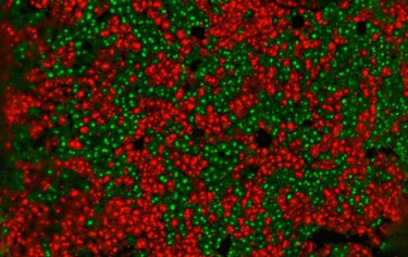
Liver section of a female mouse with X-linked GFP (green) and tdTomato (red) reporters showing patches of cells with exclusive expression of the paternally or maternally inherited X chromosome.
A central question in my laboratory is how cells count X chromosomes and initiate XCI exclusively in females. Our work showed XCI initiation is stochastic, indicating X-linked activators (Monkhorst 2008) counterbalanced by autosomal XCI-inhibitors, many of them pluripotency factors that repress Xist, linking pluripotency loss to XCI. We identified X-encoded RNF12 as an activator (Jonkers and Barakat 2009): this E3 ligase drives dose-dependent degradation of the pluripotency factor REX1 (Gontan 2012), which otherwise inhibits Xist. Because differentiating female cells retain two active Rnf12 copies, REX1 breakdown is more prominent, driving female-specific initiation. The REX1-RNF12 axis links female-specific XCI to pluripotency loss, but further work pointed to additional activators (Barakat 2015, Gontan 2018). A recent CRISPR/Cas9 screen of large X-chromosomal regions revealed HCFC1, a COMPASS member recruited by YY1 to Xist regulatory regions, increasing H3K4me3 and activating transcription. YY1 also recruits Integrator genome-wide, including at Xist, showing XCI mechanisms inform gene regulation broadly. Current work aims to identify novel XCI-activators, define how they direct female-exclusive XCI, and translate findings to humans.
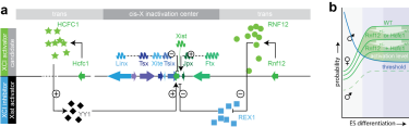
XCI regulatory network. a) Xist regulatory network of known Xist cis-regulatory genes (arrows depict genes: positive regulators of Xist in green, negative in blue), and trans-acting factors (X-linked XCI-activators in green, autosomal encoded Xist-activators and XCI inhibitors in black and blue, respectively). Long non-coding RNA genes are indicated with waves. b) Only in female cells is the concentration of XCI activators sufficient to reach the threshold required for Xist activation. This activation level is reduced in female cells carrying heterozygous deletions of the XCI activators Rnf12 and/or Hcfc1.
Epigenetic mechanisms in stem cells and disease
The development of more than 300 cell types present in our body requires the action of complex molecular mechanisms, involving signal transduction pathways that instruct gene transcription by impacting on transcription factor networks and the epigenetic landscape. Epigenetic mechanisms play a crucial role in installing and maintaining cell fate decisions. Our interest and expertise in embryonic stem cells, which we use as a model system to study XCI, has sparked research lines involving addressing research questions related to stem cell biology. For instance, our laboratory provided critical knowledge and infrastructure to setup organoid studies which were performed in close collaboration with clinical research groups, either through ESC or iPSC differentiation to kidney or forebrain organoids or expansion of tissue into kidney or placental organoids. We recently developed ETX embryo’s through self-assembly of different stem cell lines (ESC, TSC, and XEN, Dupont 2023). These models resemble post-implantation egg cylinder in vivo embryo’s providing a powerful model system to study embryonic development in vitro as well as XCI. To better understand cell state changes in development and cell differentiation we developed the DCM time machine (DCM-TM) technology (Boers, Boers and Tan 2023). DCM-TM enables the reconstruction of gene and enhancer activity maps of the past. DCM-TM utilizes an inducible DCM-RNA polymerase subunit-b fusion protein to label active genes and enhancers with a bacterial methylation mark. This mark does not interfere with transcription and is stably propagated in S-phase. We applied DCM-TM to study intestinal homeostasis, revealing new insights into cell signalling and chromatin and transcription factor activity dynamics in stem cell differentiation. DCM-TM is a versatile tool that can be applied to follow cell differentiation, embryonic development and tissue regeneration, identifying temporal maps of transcription factor networks and signal transduction pathways that can be utilized to improve stem cell expansion and cell differentiation models (van Leeuwen and Tan 2026).
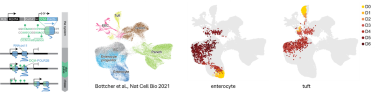
The DCM-TM technology. DCM-TM experimental setup (left), where DCM labelling of gene bodies is induced and maintained over multiple cell divisions. The right panels show annotation of cell types in a scRNA-seq UMAP and projection of cell state changes observed with DCM-TM with enterocyte and tuft cell viewpoints (in days chase).
Translation of knowledge and technology to the clinic.
The focus of our laboratory is on developing mechanistic insights into gene regulation and translating this knowledge and the technologies arising from our research into clinically relevant applications. A key example of this translational approach is the development of Methylated DNA sequencing (MeD-seq; Boers 2018; Boers 2023). MeD-seq is based on the methylation-dependent restriction enzyme LpnPI, which generates 32-base-pair DNA fragments around fully methylated target sequences. In addition to detecting DCM methylation (required for DCM-TM), MeD-seq captures approximately 50% of all methylated CpG sequences across the genome. Importantly, the technology requires very low amounts of input DNA (<2.5 ng or <500 cells), making it particularly suitable for challenging clinical samples, including laser-dissected FFPE material and cell-free DNA (cfDNA).
The combination of genome-wide methylation profiling with minimal input requirements has enabled extensive collaboration with clinicians across a broad range of disease areas. These studies have demonstrated the considerable potential of MeD-seq and, in particular, cfDNA-based applications for the minimally invasive and longitudinal assessment of disease and treatment response. Importantly, the applications of this technology extend beyond cancer diagnosis and treatment monitoring. Our collaborative studies have demonstrated its potential for the early detection of frontotemporal dementia (FTD), as well as for monitoring placental development and identifying preeclampsia at an early stage (Deger 2021; Giannini 2024; van Vliet 2025).
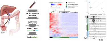
Clinical application of MeD-seq: cfDNA of controls and pre- and post-operative patients with metastasized colon cancer to the liver were subjected to MeD-seq showing loss of the cancer specific DNA methylation profile in post-operative patients (middle panel), that can be quantified (upper right panel). EYA4 is a marker specifically methylated in colon cancer (right bottom panel, collaboration with Saskia Wilting and John Martens, department of Medical Oncology).