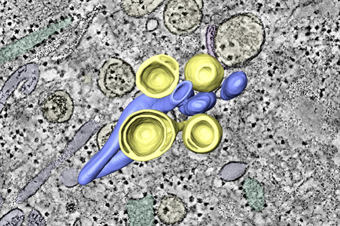
Like all positive-sense RNA viruses, picornaviruses induce the rearrangement of host-cell membranes to form viral replication organelles that support viral RNA synthesis. Here, in collaboration with Utrecht University and the LUMC Medical Microbiology department, we investigate the architecture and biogenesis of the replication organelles induced by a model cardiovirus, and compare them with those of enteroviruses, members of a distantly-related and well-characterized picornavirus genus. Using electron tomography we unravel the formation of cardiovirus replication organelles from the endoplasmic reticulum and their transformation along infection. We show that PI4P, a critical lipid for cardiovirus replication, can be spared for the formation of normal replication organelles that accommodate viral RNA synthesis. Our data reveals striking similarities with enteroviruses that may well extend to the picornavirus family at large but that so far are unique among positive-sense RNA viruses.