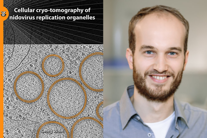
In his thesis, Georg Wolff analyzed the replication organelles induced by corona- and arteriviruses using cellular electron cryo-tomography (cryo-ET). Cellular cryo-ET is a young technique, which makes use of focused ion beam (FIB) milling to generate 100-300 nm thin cryo-lamellae from thick regions of cells that are in this way accessible to be imaged by high-resolution cryo-EM. However, this workflow is time-consuming and error-prone. Part of the thesis describes an improved sample-preparation strategy that successfully increased the throughput of this workflow. This method was applied on cells infected by coronaviruses unveiling a novel pore complex that seems to shuttle viral RNA across the double-membranes of the viral replication organelles. It was found that this molecular pore are (partially) formed by a large transmembrane viral proteins. Further work revealed that DMV-spanning molecular pores appear to be a central component in the replication cycle of both corona- and arteriviruses, two distantly related virus families united in the Nidovirales order.
Georg Wolff obtained his doctorate cum laude
Georg Wolff obtained his PhD thesis on the “Cellular cryo-tomography of nidovirus replication organelles” cum laude. The work was conducted under the supervision of dr. Montse Barcena, Prof.dr. A.J.Koster and Prof.dr. E.J.Snijder.
