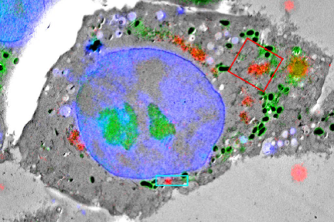
Correlative light-lectron microscopy (CLEM) combines the high spatial resolution of transmission electron microscopy (TEM) with the capability of fluorescence light microscopy (FLM) to locate rare or transient cellular events within a large field of view. CLEM is etherefore a powerful technique to study cellular processes. Aligning images derived from both imaging modalities is a prerequisite to correlate the two microscopy data sets, and poor alignment can limit interpretability of the data. Here, we describe how uranyl acetate, a commonly-used contrast agent for TEM, can be induced to fluoresce brightly at cryogenic temperatures (−195°C) and imaged by cryoFLM using standard filter sets.
DOI: 10.1038/s41598-017-10905-x
