A new publication in Nature Regenerative Medicine from Marije Koning from the group of prof. dr. Ton Rabelink showed that human induced pluripotent stem cell-derived kidney organoids have potential for disease modeling and to be developed into clinically transplantable auxiliary tissue.
Due to the lack a functional vasculature, the sparse endogenous endothelial cells that are lost upon prolonged culture in vitro, limit maturation and applicability. Intracoelomic transplantation in chicken embryos, followed by single-cell RNA sequencing and advanced imaging platforms was used to induce and study vasculogenesis in kidney organoids.
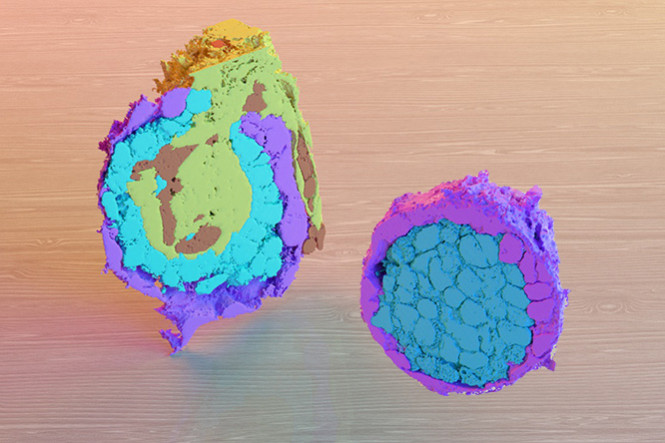
The electron microcsopy facility of the LUMC was involved in advanced volume scanning electron microscopy imaging showing the large volumes of half organoid glomeruli at nanometer resolution, flollowed by image segmentation of cells, nuclei, ER and mitochondria using artificial intelligence image segmentation.
It was shown that the expansion of human organoid-derived endothelial cells that reorganize into perfused capillaries and form a chimeric vascular network with host-derived blood vessels. Ligand-receptor analysis infers extensive potential interactions of human endothelial cells with perivascular cells upon transplantation, enabling vessel wall stabilization. Perfused glomeruli display maturation and morphogenesis to capillary loop stage. The findings demonstrate the beneficial effect of vascularization on not only epithelial cell types, but also the mesenchymal compartment, inducing the expansion of ´on target´ perivascular stromal cells, which in turn are required for further maturation and stabilization of the neo-vasculature.