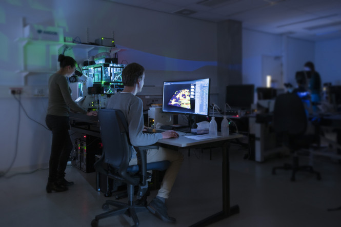
The latest addition to the light microscopy facility is a spinning-disk microscope from Andor and Leica. This new microscope expands the facilities of the TFA Light and Electron Microscopy with extremely fast and sensitive 3D imaging. But what makes spinning disk microscopy so fast and sensitive? We will have a look at two technologies that are important to understand this.
First of all, the spinning disk itself. The working principles of a spinning disk confocal microscope is very comparable to a conventional confocal laser scanning microscope (CLSM). In a confocal setup the sample is illuminated by a single point of light (a diffraction limited spot) and subsequently the fluorescent light is imaged through a pinhole in order to block all out-of-focus light. The difference between a spinning disk setup and a laser scanning confocal, is that instead of scanning the point of light over the sample using a set of scanning mirrors, the pinhole itself is on a rotating disk. The best thing about this rearrangement of optics, is that we can now efficiently multiplex the confocal principle by arranging multiple pinholes on a single spinning disk, and thereby scan multiple points simultaneously. Using a clever pinhole pattern, and by scanning the disk at 6000 rpm, we can now image an entire field-of-view in 2.5 ms. This incredible speed makes a spinning disk confocal microscope ideal for imaging live cells in 3D.
The second important thing to look at is camera technology. In a regular confocal laser scanning microscope, an image is acquired by scanning along the sample and sequentially detecting the fluorescent light using a photomultiplier tube (PMT) or hybrid detector (HyD). These detectors have an maximum efficiency of about 40%. In contrast, a spinning disk confocal uses a pixelated detector (i.e. a camera) to acquire an image instantaneously. The great thing about cameras is that they can have an efficiency up to 95%. The recently installed spinning disk system at the LUMC is equipped with a two different cameras: an EM-CCD camera with an efficiency of >95%, and a sCMOS camera with an efficiency of 82%.
Together, spinning disk microscopy and the latest camera technology make a very powerful combination to study delicate cellular processes with an incredible framerate and sensitivity.
For further information visit: Light & Electron Microscopy Facility